Ultrasound Services
PRP Diagnostic Imaging is a leading medical imaging provider specialising in a wide range of ultrasound services. Our expertise includes women’s imaging offering breast ultrasound, pelvic ultrasound, and pregnancy ultrasound including Non-Invasive Prenatal Testing (NIPT) and nuchal translucency scans. We also offer abdominal ultrasound and liver elastography, ultrasound-guided injections and biopsies, musculoskeletal ultrasound, renal ultrasound, neck/thyroid ultrasound, and vascular ultrasound.
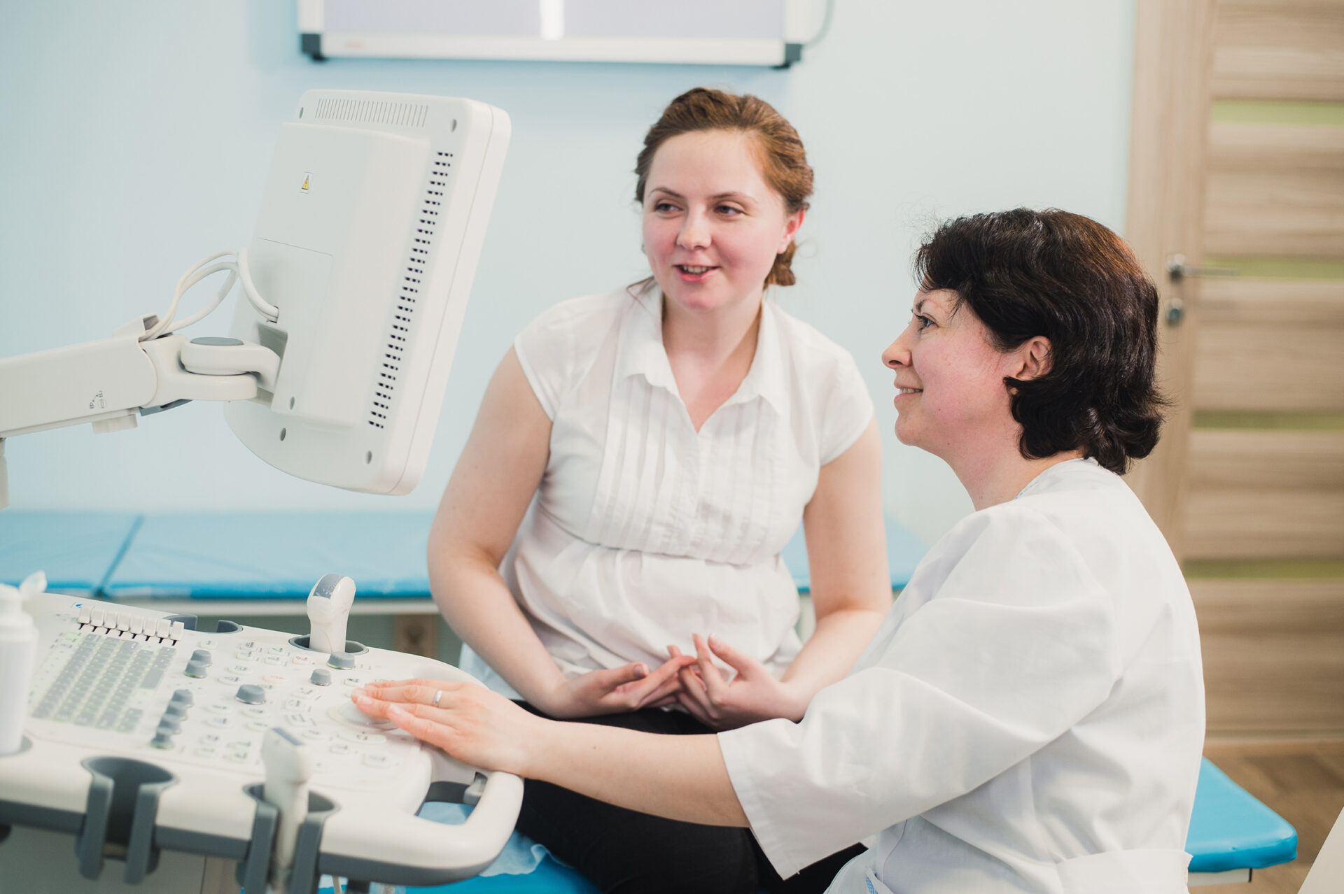
Our highly trained ultrasound sonographers will guide you through every step of your ultrasound scan to ensure you feel fully prepared for your examination and at ease.
With clinics conveniently located across New South Wales, it’s easy to schedule your ultrasound appointment at your nearest PRP practice. Book Online today.
Ultrasound Services Services available at PRP Clinics

Ankle & Foot Ultrasound
- Examines detail of ankle & foot soft tissue structures
- Takes approximately 15-30 minutes
Elbow Ultrasound
- Precise visualisation of soft tissues within the elbow
- Takes approximately 15-30 minutes
Hip Ultrasound
- Examines the hip joint and surrounding soft tissues
- Takes approximately 15-30 minutes
Knee Ultrasound
- Examines the knee joint and surrounding soft tissues
- Takes approximately 15-30 minutes
Liver Elastography Ultrasound
- Used to measure and classify liver stiffness
- Replaces liver biopsy, making it painless, safe and accurate
- Takes roughly 30 minutes
Musculoskeletal Ultrasound
- Includes muscles, tendons, joints, ligaments and soft tissues
- Takes approximately 15-30 minutes
- No preparation
Non-Invasive Prenatal Testing (NIPT)
- Prenatal screening test for common chromosomal abnormalities
- All testing & data analysis is performed in Australia.
Nuchal Translucency Scan
- Tests for a high risk of having a baby with chromosomal abnormalities
- Book at 12 weeks 4 days preferably
Pelvic Ultrasound
- Allows visualization of female pelvic organs & structures
- Requires drinking 750mls of water
- Approx 30 minutes
Pregnancy Ultrasound
- Images the unborn baby and pelvic organs
- Takes approximately 15-60 minutes
Shoulder Ultrasound
- Examines the shoulder joint and surrounding soft tissues
- Takes approximately 15-30 minutes
Ultrasound
- Images the soft tissues of your body
- Typically requires 6 hour fast
- Approx 30-60 minutes
Ultrasound Guided Injections
- Heightens the accuracy of needle placement, improving effectiveness
- Takes approximately 15-30 minutes
Vascular Ultrasound
- Images arteries or veins
- Evaluates blood flow of vessels
- Takes 30-90 minutes
Wrist & Hand Ultrasound
- Examines the wrist or hand and surrounding soft tissues
- Takes approximately 15-30 minutes
What You Need To Know About Ultrasound Procedures

Bring Necessary Documents
Please bring your referral, Medicare, and Pensioner Health Care Cards. Any previous imaging related to the region being examined is also required for comparison and accurate assessment.
Instructions
Specific preparation instructions will be provided when you book your appointment, as they vary based on the type of ultrasound.
For abdominal ultrasound, a fasting period of approximately 6 hours is typically required. Medications may be taken with a sip of water.
For renal/pelvic ultrasound, you will be instructed to empty your bladder, drink water, and then hold it for 1-2 hours before the scan.
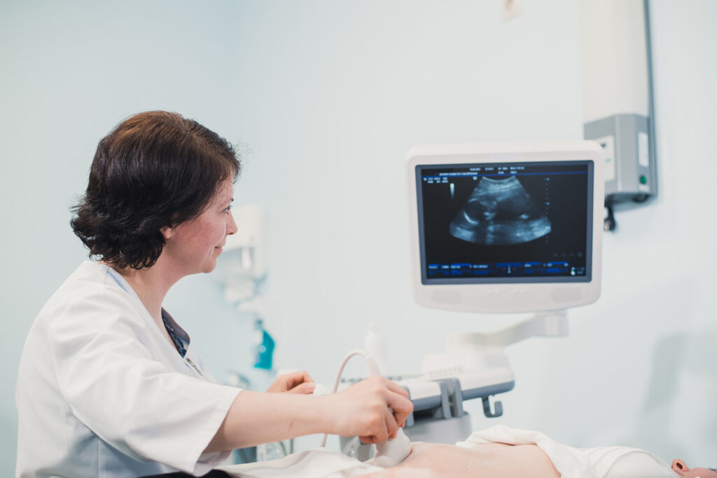
What happens during the procedure
You may be asked to change into a gown. A clear jelly is applied to the skin and a small plastic probe is moved over the skin above the area being examined. Multiple images are taken throughout the exam.
The sonographer will be with you throughout the scan and will usually ask questions about your medical history and the reason for the examination so that the optimal examination for your problem will be done.
To get the best images can be very challenging and requires considerable time and concentration by the sonographer. With consent, an internal examination may be performed during a female pelvic ultrasound.
The radiologist may need to see you to get more information or to scan to check some findings.
How long does it take?
An ultrasound takes approx 30-60 minutes, depending on the complexity.
After Your Ultrasound Procedure
There are no restrictions after having an ultrasound.
After your examination, the most pertinent images from your study will be available on the myPRP patient portal. A report, along with the images, will be sent directly to your referring doctor. PRP will store digital copies of all studies on our secure database for comparison with any future examinations.
It is important that you return to your doctor with your examination results. Whether they are normal or abnormal, your doctor needs to know promptly so that a management plan can be formulated.


What Our Patients Say About PRP
I would just like to thank the staff and Doctors, especially the sonographer who conducted my ultrasound yesterday. They were very professional and made an uncomfortable problem (for me) an easy and hassle free examination. Please pass on my gratitude.
Commonly Asked Questions About Ultrasound Services

Bulk billing is available for some services at certain PRP locations. You will be notified of any related fees or payment required when making a booking, or you can contact PRP for more information.
After your appointment, images are available to you via the myPRP patient portal. The duration for receiving the radiologist's report can vary depending on the specific examination and clinical urgency. For more detailed information, it's best to consult with the staff at your PRP clinic.
PRP ensures all patients are informed of the total cost of their exam prior to its commencement. When settling your account in full at the conclusion of your appointment, for eligible services, PRP offers the convenience of electronically submitting your account to Medicare, ensuring your rebate is deposited into your nominated bank account within 48 hours.
Ultrasound services at PRP typically require an appointment. It's advisable to schedule an appointment for ultrasound scans to ensure availability.
You can schedule your ultrasound appointment by contacting your nearest PRP clinic directly or by booking online through PRP's website. The online booking system allows you to select your preferred location and appointment time.